The mistaken claims that our faint magnetic fields can’t affect the brain ignore the evidence – a Blog by Dr. Michael Persinger.
Question: Is there any truth to the claim that your magnetic fields cannot influence the brain?

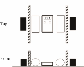
Answer: No. Recently, a colleague and I performed an experiment using three materials, each three times as dense and thick as the human skull (wood, saline solution or duct metal) to demonstrate that there is no validity to claims that weak, time-varying magnetic fields applied in this manner are eliminated or significantly attenuated (weakened) by the human skull. The result was straightforward: The fields were not attenuated (weakened) in any way.
Persinger, Michael A., and Kevin S. Saroka. “Minimum Attenuation of Physiologically-Patterned, 1 µTesla Magnetic Fields through Simulated Skull and Cerebral Space.” Journal of Electromagnetic Analysis and Applications 5.04 (2013): 151.
The contention that magnetic fields cannot influence the brain is based on a fallacious interpretation of TMS (Transcranial Magnetic Stimulation), which uses magnetic fields strong enough to depolarize neurons. Typically, these fields are a million times stronger than the kind that surround stereo headphones. This “brute force” approach has several clinical applications. Critics claim that neural stimulation employing fields with lower strengths can’t have any effect. A brief look at the applicable laws of physics and laboratory evidence shows us that this simply isn’t the case.
It should be understood that any contention that magnetic fields cannot penetrate the head are contrary to the laws of physics, which tell us that the head cannot act as a magnetic insulator, because these same laws exclude the existence of magnetic insulation. This has to do with one of Maxwell’s Equations (del dot B = 0). All magnetic field lines must terminate on the opposite pole. Because of this, there is no way to stop all of them; they must all find a way to return the magnetic field lines back to an opposite pole.
This is how it is explained in the theories of physics. When we examine the question empirically, we find that there is a substantial body of evidence showing that weak magnetic fields do penetrate the head, and that they can also influence brain activity. Let me address how this happens.
In classical physics, a changing magnetic field produces an electric field and an electric current. The amount of current depends upon the conductivity of the substance, whether its a copper wire or our brain tissues. That’s how TMS works. However, there is also magnetic energy.
When we apply our magnetic fields, with strengths a million times less than TMS, the energy within the volume of the person’s brain is about a nanoJoule (one billionth of a Joule) per second. When we average this out over the about 100 billion neurons (and their support cells) in the human brain, that works out to about 10^-20 Joules per cell each second. The number is a decimal point followed by 19 zeros and then a 1. This may seem very, very small. Actually, it matches the amount of energy involved when a single nerve cell produces one action potential that contributes to our present-time subjective experience. Moreover, a change in the activity of one neuron can alter the state of the entire brain (Cheng-Yu, 2009) This small quantity of energy is also the same as the amount that binds chemicals to cells through receptors.
However, the values can be enhanced. Our brains are richly populated with crystalline magnetite, containing 5 million such crystals per gram (Kirschvink, 1992 A). They appear in chains (“magnetosomes”). In the vernacular, our fields work because these chained crystals move in response, and because the information encoded in their movement (coming from our signals); their “patterns”, interacts with the magnetic fields that appear as a consequence of the brain’s electrical activity, a “field to field” effect. Imagine the sun has a storm, making it’s magnetic field pulse slowly. Here, on the earth, we would have geomagnetic storms, as pulses from the sun’s stormy field are added to that of our planet. We have found the same field that produced the sensed presence works by very specific channels within membranes that allow calcium to enter the cell (Buckner et al, 2015). The timing of the point durations that compose the specific field pattern must be precise or there is no effect.
One of the pioneers in biological aspects of magnetic fields, Joseph Kirshvink (1992 B) wrote: “A simple calculation shows that magnetosomes [chains of magnetic particles] moving in response to earth-strength ELF fields are capable of opening trans-membrane ion channels, in a fashion similar to those predicted by ionic resonance models. Hence, the presence of trace levels of biogenic [produced or brought about by living organisms] magnetite in virtually all human tissues examined suggests that similar biophysical processes may explain a variety of weak field ELF bioeffects”.
The magnetic fields that surround stereo headphones are in the same range, but are not embedded with neural information.
The reader can see 10 examples of magnetic stimulation studies below. Only independent studies are listed. The magnetic stimulation reported in them run from a quarter of the field strengths used in TMS (1 Tesla) to less than a millionth of that value. Each listing displays:
- The unit of magnetic field measurement used in the research publications.
- The equivalent field strength in milligauss (mG), so that the same unit of measurement can be seen for all the cited studies.
- The percent of the fields employed in TMS.
- A brief summary of each result.
- A link to the publication.
These are displayed below in descending order of field strength, and they range from one quarter (25%) to five ten-billionths (5 x 10 -13%) of the field strength used in TMS.
Wieraszko (2000) used a 2.5 milliTesla field (= 25,000 mG, which equals 0.25% of TMS) to exert effects on spikes from hippocampal slices in vitro:
Dobson, Jon, et al. (2000) used a 1.8 milliTesla field (= 18,000 mG, or 0.18% of the fields strengths used in TMS) to enhanced and suppress interictal epileptiform activity in temporal lobe epileptics.
Thomas (et al.), 2007 used a 400 microTesla magnetic field (=4,000 mG which equals 0.04% of the fields used in TMS) for pain reduction in patients with fibromyalgia.
Huesser, (et al.) 1997 used a 0.1 microTesla magnetic field (= 1000 mG , which equals 0.01% of the fields used in TMS) to cause changes in EEG parameters.
Marino (et al., 2004) used a 1 Gauss magnetic field (= 1000 mG, which equals 0.01% of the fields used in TMS) to cause changes in EEG readings during presentation of Magnetic fields
Carrubba et al., (2008) used a 2 Gauss magnetic field (= 2000 mG, which equals 0.02% of the field strengths used in TMS) to elicit magnetosensory evoked potentials.
Note: The same researcher also found EEG activation in response to magnetic fields with 1 Gauss field strengths (0.01% of the field strengths used in TMS.
Brendel et al., (2000) used an 86 microTesla magnetic field (= 860 mG or 0.0086% of the field strengths used in TMS) to elicit melatonin suppression following in vitro pineal gland exposure to magnetic fields.
Bell et al. (2007) used a 0.78 Gauss magnetic field (=780 mG or 0.0078% of the fields used in TMS) to induce increased EEG activity at two or more frequencies.
Vorobyov, et al., (1998) used a 20.9 microTesla magnetic field (=209 mG or 0.0029% of the field strengths used in TMS) to influence EEG differences in rats.
More evidence (2009).
Tinoco & Ortiz (2014) used a 1 microTesla magnetic field (=10 mG or 0.0001% of the fields strengths used in TMS) to replicate one of Persinger’s published God Helmet effects.
Jacobson (1994) used a 5 picoTesla magnetic field (= 0.00005 mG or 0.000000000005% of the field strengths used in TMS), and observed a direct correlation of melatonin production with magnetic field stimulation.
Sandyk, (1999) “picoTesla range” used 500 picoTesla (=0.005 milligauss or 0.00000000005% of the field strengths used in TMS) magnetic fields improve olfactory function in Parkinson’s disease.
NOTE: Sandyk has published scores of case histories documenting the effects of picoTesla range magnetic fields on humans, including MS and Parkinson’s.
I hope this blog will clarify that the magnetic fields we utilize in the God Helmet can indeed affect brain activity, and that claims to the contrary contradict the laws of physics and are made without examination of the evidence.
Dr. Michael A. Persinger (Passed away August 14, 2018)
Full Professor
Behavioural Neuroscience, Biomolecular Sciences and Human Studies
Departments of Psychology and Biology
Laurentian University,
Sudbury, Ontario, Canada P3E 2C6
Email questions to: brainsci@jps.net
NOTE: This blog is hosted by a colleague.
REFERENCES
Cheng-yu, T. Li, Mu-ming Poo, and Yang Dan. “Burst spiking of a single cortical neuron modifies global brain state.” Science 324.5927 (2009): 643-646.
Kirschivink, Joseph L., Kobayashi-Kisshvink, Atsuko & Woodford, Barbera J. “Magnetite biomineralization in the Human Brain”, Proceedings of the National Academy of Science 1992 (A), 89 7683-7687
Kirschvink, Joseph L., et al. “Magnetite in human tissues: a mechanism for the biological effects of weak ELF magnetic fields.” Bioelectromagnetics 13.S1 1992 (B): 101-113.
Buckner CA, Buckner AL, Koren SA, Persinger MA, Lafrenie RM (2015) Inhibition of Cancer Cell Growth by Exposure to a Specific Time-Varying Electromagnetic Field Involves T-Type Calcium Channels. PLoS ONE 10(4): e0124136. doi:10.1371/journal.pone.0124136

One Reply to “”