The God Helmet’s Weak Fields are Sufficient to Influence Brain Activity – A Blog By Dr. M. A. Persinger

The mistaken claims that our faint magnetic fields can’t affect the brain ignore the evidence – a Blog by Dr. Michael Persinger.

Question: Is there any truth to the claim that your magnetic fields cannot influence the brain?

A schematic diagram of the testing apparatus for the experiment. Two wooden pine boards or duct metal were periodically inserted between the SAM-360 device and the power meter. In one condition physiological saline was placed adjacent to the power meter.
A schematic diagram of the testing apparatus for the experiment. Two wooden pine boards or duct metal were periodically inserted between the SAM-360 (vis. God Helmet) device and the power meter. In one condition physiological saline was placed adjacent to the power meter.

Answer: No.  Recently, a colleague and I performed an experiment using three materials, each three times as dense and thick as the human skull (wood, saline solution or duct metal) to demonstrate that there is no validity to claims that weak, time-varying magnetic fields applied in this manner are eliminated or significantly attenuated (weakened) by the human skull.  The result was straightforward: The fields were not attenuated (weakened) in any way.

Persinger, Michael A., and Kevin S. Saroka. “Minimum Attenuation of Physiologically-Patterned, 1 µTesla Magnetic Fields through Simulated Skull and Cerebral Space.” Journal of Electromagnetic Analysis and Applications 5.04 (2013): 151.

The contention that magnetic fields cannot influence the brain is based on a fallacious interpretation of TMS (Transcranial Magnetic Stimulation), which uses magnetic fields strong enough to depolarize neurons.  Typically, these fields are a million times stronger than the kind that surround stereo headphones.  This “brute force” approach has several clinical applications.  Critics claim that neural stimulation employing fields with lower strengths can’t have any effect.  A brief look at the applicable laws of physics and laboratory evidence shows us that this simply isn’t the case.

It should be understood that any contention that magnetic fields cannot penetrate the head are contrary to the laws of physics, which tell us that the head cannot act as a magnetic insulator, because these same laws exclude the existence of magnetic insulation.  This has to do with one of Maxwell’s Equations (del dot B = 0).  All magnetic field lines must terminate on the opposite pole. Because of this, there is no way to stop all of them; they must all find a way to return the magnetic field lines back to an opposite pole.

This is how it is explained in the theories of physics.  When we examine the question empirically, we find that there is a substantial body of evidence showing that weak magnetic fields do penetrate the head, and that they can also influence brain activity.  Let me address how this happens.

In classical physics, a changing magnetic field produces an electric field and an electric current.  The amount of current depends upon the conductivity of the substance, whether its a copper wire or our brain tissues. That’s how TMS works.  However, there is also magnetic energy.

When we apply our magnetic fields, with strengths a million times less than TMS, the energy within the volume of the person’s brain is about a nanoJoule (one billionth of a Joule) per second.  When we average this out over the about 100 billion neurons (and their support cells) in the human brain, that works out to about 10^-20 Joules per cell each second. The number is a decimal point followed by 19 zeros and then a 1. This may seem very, very small.   Actually, it matches the amount of energy involved when a single nerve cell produces one action potential that contributes to our present-time subjective experience. Moreover, a change in the activity of one neuron can alter the state of the entire brain (Cheng-Yu, 2009) This small quantity of energy is also the same as the amount that binds chemicals to cells through receptors.

However, the values can be enhanced.  Our brains are richly populated with crystalline magnetite, containing 5 million such crystals per gram (Kirschvink, 1992 A).  They appear in chains (“magnetosomes”).  In the vernacular, our fields work because these chained crystals move in response, and because the information encoded in their movement (coming from our signals); their “patterns”, interacts with the magnetic fields that appear as a consequence of the brain’s electrical activity, a “field to field” effect.  Imagine the sun has a storm, making it’s magnetic field pulse slowly.  Here, on the earth, we would have geomagnetic storms, as pulses from the sun’s stormy field are added to that of our planet. We have found the same field that produced the sensed presence works by very specific channels within membranes that allow calcium to enter the cell (Buckner et al, 2015). The timing of the point durations that compose the specific field pattern must be precise or there is no effect.

One of the pioneers in biological aspects of magnetic fields, Joseph Kirshvink (1992 B) wrote:  “A simple calculation shows that magnetosomes [chains of magnetic particles] moving in response to earth-strength ELF fields are capable of opening trans-membrane ion channels, in a fashion similar to those predicted by ionic resonance models. Hence, the presence of trace levels of biogenic [produced or brought about by living organisms] magnetite in virtually all human tissues examined suggests that similar biophysical processes may explain a variety of weak field ELF bioeffects”.

The magnetic fields that surround stereo headphones are in the same range, but are not embedded with neural information.

The reader can see 10 examples of magnetic stimulation studies below.  Only independent studies are listed.  The magnetic stimulation reported in them  run from a quarter of the field strengths used in TMS (1 Tesla) to less than a millionth of that value.  Each listing displays:

  • The unit of magnetic field measurement used in the research publications.
  • The equivalent field strength in milligauss (mG), so that the same unit of measurement can be seen for all the cited studies.
  • The percent of the fields employed in TMS.
  • A brief summary of each result.
  • A link to the publication.

These are displayed below in descending order of field strength, and they range from one quarter (25%)  to five ten-billionths (5 x 10 -13%) of the field strength used in TMS.


Wieraszko (2000) used a 2.5 milliTesla  field (= 25,000 mG, which equals 0.25% of TMS) to exert effects on spikes from hippocampal slices in vitro:

Wieraszko, A. “Dantrolene modulates the influence of steady magnetic fields on hippocampal evoked potentials in vitro.” Bioelectromagnetics 21.3 (2000): 175-182.


Dobson, Jon, et al. (2000) used a 1.8 milliTesla  field (= 18,000 mG, or  0.18% of the fields strengths used in TMS) to enhanced  and suppress interictal epileptiform activity in temporal lobe epileptics.

Dobson, Jon, et al. “Changes in paroxysmal brainwave patterns of epileptics by weak‐field magnetic stimulation.” Bioelectromagnetics 21.2 (2000): 94-99.


Thomas (et al.), 2007 used a 400 microTesla magnetic field (=4,000 mG which equals 0.04% of the fields used in TMS)  for  pain reduction in patients with fibromyalgia.

Thomas, Alex W., (et al.) “A randomized, double-blind, placebo-controlled clinical trial using a low-frequency magnetic field in the treatment of musculoskeletal chronic pain.” Pain Research & Management: The Journal of the Canadian Pain Society 12.4 (2007): 249.


Huesser, (et al.) 1997 used a 0.1 microTesla magnetic field (= 1000 mG , which equals 0.01% of the fields used in TMS) to cause changes in EEG parameters.

Heusser, Karsten, Dieter Tellschaft, and Franz Thoss. “Influence of an alternating 3 Hz magnetic field with an induction of 0.1 microTesla on chosen parameters of the human occipital EEG.” Neuroscience letters 239.2 (1997): 57-60.


Marino (et al., 2004) used a  1 Gauss magnetic field (= 1000 mG, which equals  0.01% of the fields used in TMS) to cause changes in EEG readings  during presentation of Magnetic fields

Marino, Andrew A., et al. “Effect of low-frequency magnetic fields on brain electrical activity in human subjects.” Clinical Neurophysiology 115.5 (2004): 1195-1201.


Carrubba et al., (2008) used a  2 Gauss magnetic field (= 2000 mG, which equals 0.02% of the field strengths used in TMS) to elicit  magnetosensory evoked potentials.

Carrubba, Simona, et al. “Magnetosensory evoked potentials: consistent nonlinear phenomena.” Neuroscience research 60.1 (2008): 95-105.

Note: The same researcher also found EEG activation in response to magnetic fields with 1 Gauss field strengths (0.01% of the field strengths used in TMS.


Brendel et al., (2000) used an  86 microTesla magnetic field (= 860 mG or  0.0086% of the field strengths used in TMS) to elicit melatonin suppression following in vitro pineal gland exposure to magnetic fields.

Brendel, H., M. Niehaus, and A. Lerchl. “Direct suppressive effects of weak magnetic fields (50 Hz and 162/3 Hz) on melatonin synthesis in the pineal gland of Djungarian hamsters (Phodopus sungorus).” Journal of pineal research 29.4 (2000): 228-233.


Bell et al. (2007) used a  0.78 Gauss magnetic field  (=780 mG or 0.0078% of the fields used in TMS) to induce increased EEG activity at two or more frequencies.

Bell, Glenn B., Andrew A. Marino, and Andrew L. Chesson. “Alterations in brain electrical activity caused by magnetic fields: detecting the detection process.”
Electroencephalography and clinical Neurophysiology 83.6 (1992): 389-397.


Vorobyov, et al., (1998) used a  20.9 microTesla magnetic field  (=209 mG or  0.0029% of  the field strengths used in TMS) to influence  EEG differences in rats.

Vorobyov, Vasily Vasilievitch, et al. “Weak combined magnetic field affects basic and morphine-induced rat’s EEG.” Brain research 781.1 (1998): 182-187.

More evidence (2009).


Tinoco & Ortiz (2014) used a 1 microTesla  magnetic field  (=10 mG  or 0.0001% of the fields strengths used in TMS) to replicate one of Persinger’s published God Helmet effects.

Tinoca, Carlos A., and João PL Ortiz. “Magnetic Stimulation of the Temporal Cortex: A Partial “God Helmet” Replication Study.” Journal of Consciousness Exploration & Research 5.3 (2014).


Jacobson (1994) used a 5 picoTesla magnetic field (= 0.00005 mG or  0.000000000005% of the field strengths used in TMS), and observed a  direct correlation of melatonin production with magnetic field stimulation.

Jacobson, J. I. “Pineal-hypothalamic tract mediation of picoTesla magnetic fields in the treatment of neurological disorders.” Panminerva medica 36.4 (1994): 201-205.


Sandyk, (1999) “picoTesla range” used 500 picoTesla (=0.005 milligauss or 0.00000000005% of the field strengths used in TMS) magnetic fields improve olfactory function in Parkinson’s disease.

Sandyk, Reuven. “Treatment with AC pulsed electromagnetic fields improves olfactory function in Parkinson’s disease.” International journal of neuroscience97.3-4 (1999): 225-233.


NOTE: Sandyk has published scores of case histories documenting the effects of picoTesla range magnetic fields on humans, including MS and Parkinson’s.

I hope this blog will clarify that the magnetic fields we utilize in the God Helmet can indeed affect brain activity, and that claims to the contrary contradict the laws of physics and are made without examination of the evidence.

Dr. Michael A. Persinger  (Passed away August 14, 2018)
Full Professor
Behavioural Neuroscience, Biomolecular Sciences and Human Studies
Departments of Psychology and Biology
Laurentian University,
Sudbury, Ontario, Canada P3E 2C6
Email questions to: brainsci@jps.net
NOTE: This blog is hosted by a colleague.

REFERENCES

Cheng-yu, T. Li, Mu-ming Poo, and Yang Dan. “Burst spiking of a single cortical neuron modifies global brain state.” Science 324.5927 (2009): 643-646.

Kirschivink, Joseph L., Kobayashi-Kisshvink, Atsuko & Woodford, Barbera J. “Magnetite biomineralization in the Human Brain”, Proceedings of the National Academy of Science 1992 (A), 89 7683-7687

Kirschvink, Joseph L., et al. “Magnetite in human tissues: a mechanism for the biological effects of weak ELF magnetic fields.” Bioelectromagnetics 13.S1 1992 (B): 101-113.

Buckner CA, Buckner AL, Koren SA, Persinger MA, Lafrenie RM (2015) Inhibition of Cancer Cell Growth by Exposure to a Specific Time-Varying Electromagnetic Field Involves T-Type Calcium Channels. PLoS ONE 10(4): e0124136. doi:10.1371/journal.pone.0124136

We Do Not Allow Suggestion or Suggestibility to Influence our Lab Results. – A Blog by Dr. M.A. Persinger

We take every measure to ensure that our subjects are not exposed to suggestions.

Our laboratory results are not due to suggestion or suggestibility – A blog by Dr. Michael A. Persinger.

We apply several procedures to guarantee that our subjects are not exposed to suggestions, have no expectations, so that our results are not influenced by subject suggestibility.

These procedures and analytical methods rule out suggestion as an explanation for the effects we have observed in our experiments.

Question: How do you ensure that subjects are not inadvertently given suggestions as to the purpose of your experiments and how do you respond to the claim that your results are due to suggestibility?

Answer: Human beings are remarkably sensitive to subtle cues in their environment. For example, specific areas of the human brain respond to alterations in structures of sentences while a person is reading even though the person is not “aware” of the change in sentence structure (Bern, 1997).

Thirty years ago when we were interested in the “subjective narrative” of people sitting in the dark in a quiet chamber we found that the music, e.g., a Gregorian chant or the bars from the movie Close Encounters of the Third Kind, compared to sitting in silence, affected the content of the “spontaneous” themes.  Sitting in silent darkness without previously hearing any music generated more “death” images, the pre-darkness listening to Gregorian chants was associated with more religious images and the movie score was associated with space themes.  We could influence what the subjects thought about by “priming” them with music with clear connotations.

Context is also important (Persinger, 1989; 1992). In a placebo-controlled experiment, we applied our magnetic signals, and immediately afterwards, the subjects listened to an ambiguous narrative (story) about a young boy who had night time anomalous experiences (“The Billy Story”) .  These were epileptic in origin, although this was never stated.  After the story and the stimulation was complete, we asked to subjects to listen to a brief story about either alien abduction or early sexual abuse.  The story was provided without any explanation.  The subjects were then asked to interpret the story about the boy’s night time experiences both immediately and a few days after.  We found that the subjects who heard the story about early sexual abuse interpreted the “Billy” story as one of sexual abuse, and the subjects who heard the story about alien abductions interpreted it as being about an interaction with aliens.   (Dittburner and Persinger, 1993; O’Gorman and Persinger, 1998). In this way, we verified how easily pseudomemories or false memories can be produced (Persinger, 1992; Healey and Persinger, 2001).

My critics who attribute God Helmet experiences to suggestibility have never directly tested this hypothesis. We have tested this potential confounding effect by psychometric inferences which are highly correlated with hypnotizability, such as the Wilson-Barber Imaginings Scale. We also have several experiments where we measured hypnotizibility directly with the Spiegel and Spiegel scale where the experimenter interacts with the subject directly (Ross and Persinger, 1987). The latter was administered after the exit questionnaire containing 20 questions about their experiences.

Interestingly, the last item in this questionnaire asks if the red light changed intensity even though for most studies there was no red light. Suggestible or highly hypnotizable individuals frequently respond “yes” to this item. However, even when the many suggestibility measures were taken into account during the statistical analyses, the sensed presence reports still occurred primarily when the “God Experience” protocol (Persinger, 2001) was used.

Dr. Linda St-Pierre and I explained this in our 2006 paper in the International Journal of Neuroscience, but online skeptics seem not to have read it.   Finally I reiterate that the volunteers do not know if they will receive a magnetic field or which field it might be and they are always told they are participating in a relaxation study.

We use questionnaires with our student subjects.  The questionnaires are applied at least six weeks before these students participate, and these are only some of the questionnaires they students fill out as they study psychological data gathering.  This gives them first hand experience with methods of gathering psychological data.  They have no idea that the questions have anything to do with the experiment.  The data is gathered under “blind” conditions.

We do not decorate our lab with any spiritual or religious imagery, and the researchers don’t discuss the specifics of the experiment with subjects until after all data have been gathered.

One experiment made it clear that our effects were not coming from suggestion.  After experimenting for several years with a burst-firing pattern, and seeing repeated results telling us that it generated higher pleasantness ratings when applied over the left hemisphere (see example; Persinger & Healey, 2002), we began to investigate another signal (derived from hippocampal tissues during long-term potentiation).  In that case, and in a few since, we have seen that this signal is more pleasant over the right hemisphere (Persinger, et al., 1994).  If our effects were due to suggestion, these results should have been the same in all cases, but they weren’t.  We’ve also had many other results (for example, with lab rats) that simply cannot be explained by suggestion or suggestibility.

I hope this blog clarifies that we are aware of experimental factors that allow suggestions and suggestibility to confound experimental results, and that we take all necessary steps to prevent their occurrence.

Dr. Michael A. Persinger
Full Professor
Behavioural Neuroscience, Biomolecular Sciences and Human Studies
Departments of Psychology and Biology
Laurentian University,
Sudbury, Ontario, Canada P3E 2C6
Email: mpersinger@laurentian.ca and drpersinger@neurocog.ca
NOTE: This blog is hosted by a colleague.


REFERENCES:

Berns, Gregory S., Jonathan D. Cohen, and Mark A. Mintun. “Brain regions responsive to novelty in the absence of awareness.” Science 276.5316 (1997): 1272-1275.
https://pubmed.ncbi.nlm.nih.gov/9157889/


Dittburner, T.-L. and Persinger, M. A. (1993). Intensity of amnesia during hypnosis is positively correlated with estimated prevalence of sexual abuse and alien abductions: implications for false memory syndrome. Perceptual and Motor Skills, 77, 895-898.
https://pubmed.ncbi.nlm.nih.gov/8284172/


Healey, F. and Persinger, M. A. (2001). Experimental production of illusory (false) memories in reconstructions of narratives: effect size and potential mediation by right hemispheric stimulation from complex, weak magnetic fields. International Journal of Neuroscience, 106, 195-207.
https://pubmed.ncbi.nlm.nih.gov/11264920/


O’Gorman, K. A. and Persinger, M. A. (1998). Hypnotic Induction profiles, contextual innuendo and delayed intrusion errors for a narrative: searching for mediating variables. Perceptual and Motor Skills, 87, 587-593.
https://pubmed.ncbi.nlm.nih.gov/9842607/


Persinger, M. A. (1989). Geophysical variables and behavior: LV. Predicting the details of visitor experiences and the personality of experients. Perceptual and Motor Skills, 68, 55-65.
https://pubmed.ncbi.nlm.nih.gov/2648314/


Persinger, M. A. (1992). Neuropsychological profiles of adults who report “suddenly remembering” of early childhood memories: implications for claims of sexual abuse and alien visitations/abduction experiences. Perceptual and Motor Skills, 75, 259-266.
https://pubmed.ncbi.nlm.nih.gov/1528678/


Persinger, M. A. (1996). Subjective pseudocyesis in normal woman who exhibit enhanced imaginings and elevated indicators of electrical lability within the temporal lobes: implications for the “Missing Embryo Syndrome”. Social Behavior and Personality, 24, 101-112.
https://pubmed.ncbi.nlm.nih.gov/1852557/


Persinger, Michael A. “The neuropsychiatry of paranormal experiences.” The Journal of neuropsychiatry and clinical neurosciences 13.4 (2001): 515-524..

https://neuro.psychiatryonline.org/doi/10.1176/jnp.13.4.515


Ross, J. and Persinger, M. A. (1987). Positive correlations between temporal lobe signs and hypnosis induction profiles: a replication. Perceptual and Motor Skills, 64, 828-830.
https://pubmed.ncbi.nlm.nih.gov/3601605/


St-Pierre, L. S. and Persinger, M. A. (2006). Experimental facilitation of the sensed presence is predicted by specific patterns of applied magnetic fields not suggestibility: re-analysis of 19 experiments. International Journal of Neuroscience, 116, 1-8.
https://pubmed.ncbi.nlm.nih.gov/16861170/


Spiegel, H. and Spiegel, D. (1978). Trance and treatment: clinical uses of hypnosis. New York: Basic Books.
https://psycnet.apa.org/record/2004-13877-000


Persinger, Michael A., and Faye Healey. “Experimental facilitation of the sensed presence: Possible intercalation between the hemispheres induced by complex magnetic fields.” The Journal of nervous and mental disease 190.8 (2002): 533-541.
https://pubmed.ncbi.nlm.nih.gov/12193838/


Persinger, Michael A., Pauline M. Richards, and Stanley A. Koren. “Differential ratings of pleasantness following right and left hemispheric application of low energy magnetic fields that stimulate long-term potentiation.” International journal of neuroscience 79.3-4 (1994): 191-197.
https://pubmed.ncbi.nlm.nih.gov/7744561/

Our results can’t be attributed to suggestion. – A blog By Dr. Michael Persinger

Shiva_God_HelmetThe results of our experiments using complex magnetic stimulation cannot be attributed to experimenter suggestion or subject suggestibility. – A Blog by Dr. Michael Persinger.

Question: What results have you had that cannot be explained by suggestion?

Answer: We have seen many effects from our complex signals that cannot be explained by suggestion or suggestibility.  Some of them have been done with people, rats, worms and living cells maintained (cultured) in petri dishes, and you can’t influence living cells (in vitrio) through suggestion.

One very clear illustration is seen in our study that found that our magnetic field stimulation actually enhanced hypnotizability (Tiller, 1994) with a pulsed field, which we replicated two years later (Healey, 1996) with a burst-firing pattern.    We took measures of the suggestibility of each subject using an established instrument for its measurement as part of these experiments (Spiegel, 1978).  Altering suggestibility through suggestion is a highly improbable scenario, so the results of these studies ruled it out as an explanation for our magnetic stimulation effects in humans approximately twenty years ago.

For many of our experiments that created the sensed presence, we measured suggestibility directly using a well-established protocol (Spiegel, 1978).  We found that analyzing the data for the person’s hypnotizability score did not reduce the intensity of the sensed presence produced by the specific magnetic field pattern. The key paper was called Experimental facilitation of the sensed presence is predicted by specific patterns of applied magnetic fields not by suggestibility: re-analysis of 19 experiments”. It was published in the International Journal of Neuroscience (St-Pierre, LS, 2006).

We have observed clinical effects that cannot be explained by suggestion. For example, Baker-Price (1996) found differential changes in the EEG patterns of patients who had sustained head injuries, depending upon the specific neural location where the field was applied.  The experiment also found a significant improvement of depression and a reduction of phobias.  We did a replication and electroencephalographic validation of this experiment in 2003 (Baker-Price, 2003), with very similar results and follow-up six weeks after the experiment was completed.  Tsang et. al. (2009) showed clearly with relatively crude psychometric measurements that infer emotional profiles that different patterned fields produced different mood states.  One of them improved mood and vigour, compared to the sham-field.  Attributing these results to patient suggestibility leads inevitably to the conclusion that  depression can be treated through suggestion, a conclusion not supported by any evidence nor predicted by any hypotheses.

We have also shown (Corradini et al.  2013) memory enhancement and  clear changes in delta and theta power over the temporal lobes from magnetic field stimulation (and more accurate brain wave measures, such as QEEG), using a completely separate method for producing the God Helmet’s quiet magnetic signals.  Eliciting the same effects with both different hardware and software also allows confirmation that our results that cannot be attributed to hardware artifacts.

However, the most powerful demonstration that the electromagnetic effects are not due to suggestion is the effect upon cells. We have found in controlled studies that the same pattern using in our sensed presence experiments slows the rate of a variety of different types of cancer cells in cultures (Hu, et al., 2010, Bruckner, 2015). These fields inhibited only cancer cell growth but did not retard the growth of normal cells.  We have found this same field that produced the sensed presence works by very specific channels within membranes that allow calcium to enter the cell (Buckner et al, 2015). The timing of the point durations that compose the specific field pattern must be precise or there is no effect.

Our studies using lab rats cannot be attributed to suggestion as rats cannot be said to be “suggestible”.

Nevertheless, we carry out blind analysis of rat brain sections in our rat studies, in which the investigator does not know which brain regions may have been affected by a procedure or the magnitude of the differences predicted between the rat brains exposed to the magnetic fields and those which were not (Fournier, 2012). In rat studies investigating differences in rat behavior following stimulation with magnetic signals, the experimenter observing their behavior is kept blind to the experimental condition (Whissell, 2007, McKay, 2004, Bureau, 1994, Babik, 1992).  Our examination of microscope slides from rat subjects and controls is also done under blind conditions (Cook, 1999). We have also carried out similar procedures with worms (planarium – Dugesia sp.) (Mulligan, 2012).

The false impression that our effects come from suggestibility of our experimental participants originates from  a paper published by Granqvist et al..  That publication reported a flawed attempt at replicating our work.  In that case our neural stimulation signals were run at too high a velocity (their computer wasn’t calibrated to the software) and for too short a time (only ten minutes instead of the twenty we normally use as the minimum stimulation).   There were other issues, but these were the most significant.  Granqvist, et al. tried to explain the difference between our results and theirs by speculating that our results were due to suggestibility in our subjects. Note that Granqvist (et al.) did not actually measure their subject’s suggestibility.   Their explanation is entirely speculative and ultimately incorrect.

I hope this blog will clarify that we are fully aware of the need to prevent experimenter suggestions and that our results are not due to to suggestibility in our subjects.

Dr. Michael A. Persinger
Full Professor
Behavioural Neuroscience, Biomolecular Sciences and Human Studies
Departments of Psychology and Biology
Laurentian University,
Sudbury, Ontario, Canada P3E 2C6
Email: mpersinger@laurentian.ca and drpersinger@neurocog.ca
NOTE: This blog is hosted by a colleague.


REFERENCES

Tiller, S. G., and Michael A. Persinger. “Enhanced hypnotizability by cerebrally applied magnetic fields depends upon the order of hemispheric presentation: An anistropic effect.” International journal of neuroscience 79.3-4 (1994): 157-163.

Healey, Faye, Michael A. Persinger, and S. A. Koren. “Enhanced hypnotic suggestibility following application of burst-firing magnetic fields over the right temporoparietal lobes: A replication.” International journal of neuroscience 87.3-4 (1996): 201-207.

Spiegel, H. & Spiegel, D. (1978) Trance and treatment. Basic Books: N.Y.

Pierre, LS St, and M. A. Persinger. “Experimental facilitation of the sensed presence is predicted by the specific patterns of the applied magnetic fields, not by suggestibility: re-analyses of 19 experiments.” International Journal of Neuroscience 116.19 (2006): 1079-1096.

Baker-Price, L. A., and Michael A. Persinger. “Weak, but complex pulsed magnetic fields may reduce depression following traumatic brain injury.”  Perceptual and motor skills 83.2 (1996): 491-498.

Baker-Price, Laura, and Michael A. Persinger. “Intermittent burst-firing weak (1 microTesla) magnetic fields reduce psychometric depression in patients who sustained closed head injuries: A replication and electroencephalographic validation.” Perceptual and motor skills 96.3 (2003): 965-974.

Tsang, Eric W., Stanley A. Koren, and Michael A. Persinger. “Specific patterns of weak (1 microTesla) transcerebral complex magnetic fields differentially affect depression, fatigue, and confusion in normal volunteers.” Electromagnetic biology and medicine 28.4 (2009): 365-373.

Corradini, Paula L.; Mark W. G. Collins; Dr. Michael A. Persinger  “Facilitation of Declarative Memory and Congruent Brain States by Applications of Weak, Patterned Magnetic Fields: The Future of Memory Access?”  International Journal of Humanities and Social Science Vol. 4, No. 13; November 2014

Hu, Jing H., et al. “Growth of injected melanoma cells is suppressed by whole body exposure to specific spatial-temporal configurations of weak intensity magnetic fields.” International journal of radiation biology 86.2 (2010): 79-88.

Buckner CA, Buckner AL, Koren SA, Persinger MA, Lafrenie RM (2015) Inhibition of Cancer Cell Growth by Exposure to a Specific Time-Varying Electromagnetic Field Involves T-Type Calcium Channels. PLoS ONE 10(4): e0124136. doi:10.1371/journal.pone.0124136

Whissell, P.D. , Persinger, M.A.; “Developmental effects of perinatal exposure to extremely weak 7 Hz magnetic fields and nitric oxide modulation in the Wistar albino rat ” International Journal of Developmental Neuroscience 25 (2007) 433–439

McKay, B. E., and M. A. Persinger. “Normal spatial and contextual learning for ketamine-treated rats in the pilocarpine epilepsy model.” Pharmacology Biochemistry and Behavior 78.1 (2004): 111-119.

Bureau, Y. R. J., O. Peredery, and M. A. Persinger. “Concordance of quantitative damage within the diencephalon and telencephalon following systemic pilocarpine (380 mg/kg) or lithium (3 mEq/kg)/pilocarpine (30 mg/kg) induced seizures.” Brain Research 648.2 (1994): 265-269.

Missaghi, Babik, Pauline M. Richards, and Michael A. Persinger. “Severity of experimental allergic encephalomyelitis in rats depends upon the temporal contiguity between limbic seizures and inoculation.” Pharmacology Biochemistry and Behavior 43.4 (1992): 1081-1086.

Cook, Lisa L., and M. A. Persinger. “Infiltration of lymphocytes in the limbic brain following stimulation of subclinical cellular immunity and low dosages of lithium and a cholinergic agent.” Toxicology letters 109.1 (1999): 77-85.

Mulligan, Bryce P. , Noa Gang, Glenn H. Parker, Michael A. Persinger  “Magnetic Field Intensity/Melatonin-Molarity Interactions: Experimental Support with Planarian (Dugesia sp.) Activity for a Resonance-Like Process” Open Journal of Biophysics, 2012, 2, 137-143

Replication of God Helmet experiment and many other of our results. – a Blog by Dr. Michael A Persinger.

200px-Laurentian_University_CoA.svgMuch of our work, including with the God Helmet, has indeed been replicated. Claims to the contrary are mistaken . – A blog by Dr. M.A. Persinger.

Question: Have any God Helmet effects been replicated?  Have other results from your lab have seen replication?

Answer. Yes.  Every major discovery we have published that was considered “novel” has been replicated when the researcher followed the correct procedure.

A replication of God Helmet effects we have also reported in the literature was published in 2014 by a pair of Brazilian researchers (Tinoco, et al. 2014).  Their report very closely replicates one of our early studies (Richards & Persinger, 1993), using the same analytical method that distinguished subjects from controls.  That study concluded that it supports the contention that its results and those reported in our research publications are “attributable to the fields and their configurations, not to suggestibility”.  This refutes the claims that our results come from subject suggestibility.  These mistaken claims come from a researcher (Granqvist et al.) who didn’t use his equipment properly, so it yielded no results.  To explain his failure, he claimed that we used improper methods with suggestible subjects.  In fact, we do use placebo controlled, double-blind methods.

I and my colleagues have also answered these claims formally in publications in Neuroscience Letters, using data from 407 subjects. 

We have replied to points raised by Granqvist in debate in a publication showing that the (experimental facilitation) of the sensed presence is predicted by the specific patterns of the magnetic fields, not by suggestibility.

In addition, we’ve reviewed the methodological differences (between our methods and those used in the Swedish Research group’s flawed work) informally online.

The Granqvist et al study was not designed to be a replication. They did not use the well-known helmet but instead employed a device we made for them as a prototype so they could study PET (Positron Emission Tomography) effects. This is what they claimed they were going to do when they visited the laboratory, and that was what we had in mind when we gave them their instructions for its use. They did not follow our precise timing procedures, needed to generate the fields. The effects do not occur without this precision timing.  Granqvist et al presented the fields in a non-optimal manner, and their scores for their sham and treatment groups were the same as the scores for our sham groups.  Effective magnetic fields are sensitive to the complexity of the timing of their components, just as chemical actions depend on the exact shape of the chemicals being employed. It is like changing a component of a drug and wondering why it does not work.

Granqvist wrote: “Such weak fields are considered unable to induce currents strong enough to depolarize neurons. Thus, the mechanism through which weak complex field(s) … may work remains obscure, but the waveform of the field has been suggested to be crucial” (Granqvist, 2004).  In fact, there is nothing obscure about how the fields work in the brain.

One of the pioneers in biological aspects of magnetic fields, Joseph Kirshvink (1992 A) wrote:  “A simple calculation shows that magnetosomes [chains of magnetic particles] moving in response to earth-strength ELF fields are capable of opening trans-membrane ion channels, in a fashion similar to those predicted by ionic resonance models. Hence, the presence of trace levels of biogenic [produced or brought about by living organisms] magnetite in virtually all human tissues examined suggests that similar biophysical processes may explain a variety of weak field ELF bioeffects”.

Our brains are richly populated with crystalline magnetite, containing 5 million such crystals per gram (Kirschvink, 1992 B).  In the vernacular, our fields work because these crystals move in response and because the information encoded in their movement (coming from our signals); their “patterns”, interacts with the magnetic fields that appear as a consequence of the brain’s electrical activity, a “field to field” effect.  Imagine the sun has a storm, making it’s magnetic field pulse slowly.  Here, on the earth, we would have geomagnetic storms, as pulses from the sun’s stormy field is added to that of our planet. We have found this same field that produced the sensed presence works by very specific channels within membranes that allow calcium to enter the cell (Buckner et al, 2015). The timing of the point durations that compose the specific field pattern must be precise or there is no effect.

Notwithstanding the explanation for the effects of the fields, we also know of at least a dozen researchers who have observed measurable effects from  low-intensity magnetic fields that are significantly weaker than TMS.  These corroborate our reports of measurable bioeffects from weak magnetic fields and demonstrate that depolarizing neurons is not the only way magnetic fields can influence the brain.  We also did a study to demonstrate the permeability of the skull and cerebral space to magnetic fields (Persinger & Saroka, 2013).

Our work has been replicated and confirmed (Tinoco, 2014), which appears to resolve these issues.  We look forward to further studies in the field.

Our analgesic effects from weak applied magnetic fields, beginning in 1994 (Fleming, et al., 1994, Martin, 2004,  Gallic, 2007) were replicated by Thomas, Prato & Kavaliers in invertebrates (Thomas, 1997), rats (Shupak, 2004),  and humans (Robertson, et al., 2010).  A major review by an Italian research group (Seppia, et al., 2007) has demonstrated not only the effectiveness of these fields but their biochemical pathways.  Similar research has been done by many others (Bao, 2006, Kortekaas, 2014,).  Our work on these effects continues up to the present (Murugan, 2014).

The Tectonic Strain Theory has seen a number of replications, corroborations, and validations.  In fact, this work has been carried out  by several independent researchers.

The same is true for our work on geomagnetic influences on paranormal events and geomagnetic influences levels.

My original work (Persinger, 1969) concerning the effects of rotating magnetic fields upon fetal development in rats was replicated by Ossenkopp (1972).

My quantitative demonstration that proportion of global warming is related to solar corona expansion and trends in geomagnetic activity rather than only CO2 (Persinger, 2010) has been replicated by several researchers, including Sudhir (2012) and Wilson, (2014).

Our histopathological analysis of calcium aggregates in brain from excessive seizure damage (Lafreniere et al, 1992) was replicated by Japanese researchers (Mori, 2000).

Our LORETA measures concerning mental time travel, that is measuring memories of experiences from the past and present and pre-experiences of the imagined future (Lavallee, et al., 2010) are consistent with what many others have measured, including Irish (2013).

It seems that one researcher did not replicate one of our results, and online commentators have taken this to mean that none of them have been replicated.  This, in turn was distorted to imply that none of them could be.  I have little doubt that this chain of rumors and distortions has its basis in the way some have been offended by my work on religion.  Religious believers don’t want to accept that the brain could be instrumental in religious belief, and “new atheists” are offended by the idea that religion and religious experiences reflect processes intrinsic to the human brain, and can thus be considered as intrinsic to our species.

Anyone who has taken the time to precisely recreate the experimental or analytical conditions have replicated and extended our results.

I hope this blog will clarify that much of our work has been replicated and that we look forward to persevering in our efforts.

Dr. Michael A. Persinger
Full Professor
Behavioural Neuroscience, Biomolecular Sciences and Human Studies
Departments of Psychology and Biology
Laurentian University,
Sudbury, Ontario, Canada P3E 2C6
Email: mpersinger@laurentian.ca and drpersinger@neurocog.ca
NOTE: This blog is hosted by a colleague.


REFERENCES

St-Pierre, L.S., and M. A. Persinger. “Experimental facilitation of the sensed presence is predicted by the specific patterns of the applied magnetic fields, not by suggestibility: re-analyses of 19 experiments.” International Journal of Neuroscience 116.19 (2006): 1079-1096.
https://pubmed.ncbi.nlm.nih.gov/16861170/

Persinger, Michael A., and S. A. Koren. “A response to Granqvist et al.“Sensed presence and mystical experiences are predicted by suggestibility, not by the application of transcranial weak magnetic fields”.” Neuroscience Letters 380.3 (2005): 346-347.
https://pubmed.ncbi.nlm.nih.gov/15862915/

Tinoca, Carlos A., and João PL Ortiz. “Magnetic Stimulation of the Temporal Cortex: A Partial “God Helmet” Replication Study.” Journal of Consciousness Exploration & Research 5.3 (2014).
https://jcer.com/index.php/jcj/article/viewFile/361/386

Richards, Pauline M., Michael A. Persinger, and Stan A. Koren. “Modification of activation and evaluation properties of narratives by weak complex magnetic field patterns that simulate limbic burst firing.” International journal of Neuroscience 71.1-4 (1993): 71-85.
https://pubmed.ncbi.nlm.nih.gov/8407157/

Granqvist, Pehr, et al. “Sensed presence and mystical experiences are predicted by suggestibility, not by the application of transcranial weak complex magnetic fields.” Neuroscience Letters 379.1 (2005): 1-6.
https://pubmed.ncbi.nlm.nih.gov/15849873/

Kirschvink, Joseph L., et al. “Magnetite in human tissues: a mechanism for the biological effects of weak ELF magnetic fields.” Bioelectromagnetics 13.S1 (1992 A): 101-113.
https://pubmed.ncbi.nlm.nih.gov/1285705/

Kirschivink, Joseph L., Kobayashi-Kisshvink, Atsuko & Woodford, Barbera J. “Magnetite biomineralization in the Human Brain”, Proceedings of the National Academy of Science 1992 (B), 89 7683-7687
https://pubmed.ncbi.nlm.nih.gov/1502184/

Buckner CA, Buckner AL, Koren SA, Persinger MA, Lafrenie RM (2015) Inhibition of Cancer Cell Growth by Exposure to a Specific Time-Varying Electromagnetic Field Involves T-Type Calcium Channels. PLoS ONE 10(4): e0124136. doi:10.1371/journal.pone.0124136
https://pubmed.ncbi.nlm.nih.gov/25875081/

M. Persinger and K. Saroka, “Minimum Attenuation of Physiologically-Patterned, 1µ Tesla Magnetic Fields through Simulated Skull and Cerebral Space,” Journal of Electromagnetic Analysis and Applications, Vol. 5 No. 4, 2013, pp. 151-156.
https://www.scirp.org/html/3-9801425_29661.htm

Fleming, Jennifer L., M. A. Persinger, and Stanley A. Koren. “Magnetic pulses elevate nociceptive thresholds: Comparisons with opiate receptor compounds in normal and seizure-induced brain-damaged rats.” Electromagnetic Biology and Medicine 13.1 (1994): 67-75.
http://neurosciarchive.byethost12.com/Magnetic-pulses-elevate-nociceptive-thresholds.pdf

MartinL.J. , S.A. Koren, M.A. Persinger “Thermal analgesic effects from weak, complex magnetic fields and pharmacological interactions”  Pharmacology, Biochemistry and Behavior 78 (2004) 217–227
https://pubmed.ncbi.nlm.nih.gov/15219761/

Galic M.A. and Persinger M.A. “Lagged Association Between Geomagnetic Activity and Diminished Nocturnal Pain Thresholds in Mice”  Bioelectromagnetics 28:577, 579 (2007)
https://pubmed.ncbi.nlm.nih.gov/17657732/

Thomas, Alex W., et al. “Antinociceptive effects of a pulsed magnetic field in the land snail, Cepaea nemoralis.” Neuroscience letters 222.2 (1997): 107-110.
https://pubmed.ncbi.nlm.nih.gov/9111740/

Robertson, John A., et al. “Evidence for a dose-dependent effect of pulsed magnetic fields on pain processing.” Neuroscience letters 482.2 (2010): 160-162.
https://pubmed.ncbi.nlm.nih.gov/20643187/

Del Seppia, Cristina, et al. “Pain perception and electromagnetic fields.”Neuroscience & Biobehavioral Reviews 31.4 (2007): 619-642.
https://pubmed.ncbi.nlm.nih.gov/17374395/

Bao, Xiuqi, et al. “A possible involvement of β‐endorphin, substance P, and serotonin in rat analgesia induced by extremely low frequency magnetic field.”  Bioelectromagnetics 27.6 (2006): 467-472.
https://pubmed.ncbi.nlm.nih.gov/16622860/

Kortekaas, R., et al. “Weak field transcerebral pulsed electromagnetic fields in health care.” Antennas and Propagation (EuCAP), 2014 8th European Conference on IEEE, 2014.
https://www.infona.pl/resource/bwmeta1.element.ieee-art-000006902110

Murugan, Nirosha J. , Lukasz M. Karbowski and Michael A. Persinger “Weak burst-firing magnetic fields that produce analgesia equivalent to morphine do not initiate activation of proliferation pathways in human breast cells in culture” Integrative Cancer Science and Therapeutics, 2014 Volume 1(3): 47-50
https://pubmed.ncbi.nlm.nih.gov/15219761/

Persinger, Michael A. “Open‐field behavior in rats exposed prenatally to a low intensity‐low frequency, rotating magnetic field.” Developmental psychobiology 2.3 (1969): 168-171.
https://pubmed.ncbi.nlm.nih.gov/5407665/

Ossenkopp, Klaus P. “Maturation and open-field behavior in rats exposed prenatally to an ELF low-intensity rotating magnetic field.” Psychological Reports, Vol 30(2), Apr 1972, 371-374.
https://pubmed.ncbi.nlm.nih.gov/5024911/

Persinger, Michael A. “The cosmology of climate change: Intercorrelations between increased global temperature, carbon dioxide and geomagnetic activity.” Journal of Cosmology 8 (2010): 1957-1969.
http://neurosciarchive.byethost12.com/The cosmology of climate change.pdf

Sudhir, Joshi Indira , Christiana, Tadiparti Mary “Linkage between Cyclonic storms, Geomagnetic storms, Sunspot numbers and Climate Change”  Research Journal of Recent Sciences  Vol. 1(2), 100-103, Feb. (2012)
http://www.isca.in/rjrs/archive/v1/i2/12ISCA-RJRS-2012-029.pdf

Wilson, Robert M. “On the Relationship Between Global Land-Ocean Temperature and Various Descriptors of Solar-Geomagnetic Activity and Climate.” NASA/TP—2014–218193, NASA Marshall Space Flight Center, Huntsville, AL 62 pp, 2014.
https://ntrs.nasa.gov/api/20140006389/

Lafreniere, Ghislaine F., Oksana Peredery, and Michael A. Persinger. “Progressive accumulation of large aggregates of calcium-containing polysaccharides and basophilic debris within specific thalamic nuclei after lithium/pilocarpine-induced seizures.” Brain research bulletin 28.5 (1992): 825-830.
https://pubmed.ncbi.nlm.nih.gov/1377589/

Mori, Fumiaki, Kunikazu Tanji, and Koichi Wakabayashi. “Widespread calcium deposits, as detected using the alizarin red S technique, in the nervous system of rats treated with dimethyl mercury.” Neuropathology 20.3 (2000): 210-215.
https://pubmed.ncbi.nlm.nih.gov/11132937/

Irish, Muireann, and Olivier Piguet. “The pivotal role of semantic memory in remembering the past and imagining the future.” Frontiers in behavioral neuroscience 7 (2013).
https://pubmed.ncbi.nlm.nih.gov/23565081/

God Helmet Experiments use Blind Protocols and Placebo Controls – A Blog By Dr. M.A. Persinger

God_helmetOur God Helmet experiments employ double-blind conditions and placebo protocols – A Blog by Dr. Michael A. Persinger.

Our critics are mistaken when they claim we do not use proper controls.  We are committed to the scientific method, especially in laboratory experiments, including subject blindness, experimenter blindness, control groups, and blindness by those who analyze our data.

Question:  What are your standard double-blind and placebo controlled protocols?

Answer:  Expectancy and confirmation bias are always important variables when human beings are measuring or being measured. Most of our major experiments over the last three decades with the sensed presence were double blind.  For a placebo, we use a “sham” or absent magnetic field, which we create by disconnecting the solenoids (magnetic coils) from the signal source.  We also exploit all ways and means for ensuring that our subjects are not given any suggestions as to the purpose of the experiment.

Subject Expectations

The subjects volunteered for a relaxation study and were told (via the consent form) they might be exposed to a weak magnetic field. Four to six weeks prior to their participation, the subjects had completed intake questionnaires.  Some critics have mistakenly said that our questionnaires (which asked about some spiritual and otherworldly experiences and beliefs) were administered immediately before the experimental sessions, and that this introduced inadvertent suggestions. In fact, our standard procedure is to separate the questionnaires from the sessions by an average of a month.  They are eventually invited to participate in a “Relaxation Experiment”, so they are unaware that the questionnaires given previously have any relation to the experiment.  The subjects are kept in the experimentally blind condition.  They are not influenced by expectations when coming into our lab.  The lab itself looks like a busy workplace, and is not decorated with religious or spiritual images.

Blind Protocols

The experimenter, usually an undergraduate or graduate student, who runs the experiment is not aware of the true hypothesis or mechanism.

One summary of our work with the sensed presence is our publication: The sensed presence within experimental settings: implications for the male and female concept of self” The Journal of Psychology, 2003, 137, 5-16.
https://pubmed.ncbi.nlm.nih.gov/12661700/

Here is a very brief summary of that experiment, with its 50 male and 50 female subjects.

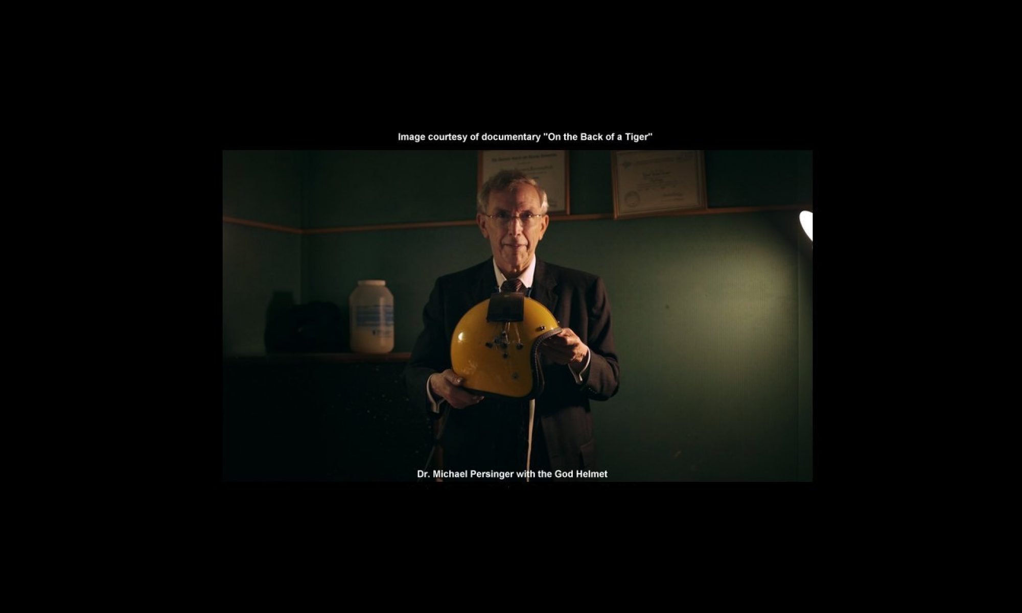
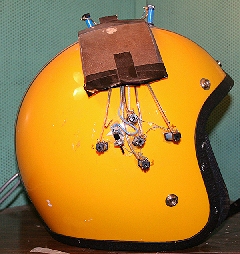
In this study, we used a signal derived from burst-firing in the amygdala, applying it over the temporal lobes via a set of four solenoids over each temporal lobe.  We rotated the signal by turning them on and off in sequence.  All of this was built into the hardware, built by Stanley Koren (The God Helmet) or coded into the software that drives it, written by Stanley Koren.  We applied the signal for 10 minutes, gave it a 5 minute break, and applied it for another 10 minutes.  This was done to avoid habituation.

We observed  double-blind conditions, as one can see in the research report:

“All participants were tested by experimenters who were not familiar with the purpose of the experiment.”
“The participants were told that the experiment was concerned with relaxation.”

Note that in some experiments, subjects were told that the experiment concerned memory.  The relaxation and memory suggestions kept the subject in an experimentally blind condition.

The experimenter ran different patterns of magnetic fields created by Professor Stan Koren and me.  Once the results were collected they were analyzed routinely by SPSS  (statistical analysis) software.

Women reported more frequent experiences of a sensed presence than men did , and men were more likely than women to consider these experiences as “intrusions” from extrapersonal or ego-alien sources. Both effects were predicted by one of our hypotheses (vectoral hemisphericity) and the known neurologically-based cognitive differences between right-handed men and right-handed women.

The point here is that we do in fact use double-blind conditions, and claims to the contrary are simply not true.  Other examples, referenced here, include Richards (1992) Persinger (1994), Healey (1996), Persinger (1999), Persinger, (2002) Booth (2005), Tiller (2002), Corradini, (2014).  I have emphasized this in my response to Pehr Granqvist (who made critical technical errors with our equipment, and alleged that our results were due to improper blinding and subject suggestibility), as follows:

“In all of our major studies, involving more than 400 subjects, during the last 20 years the subjects were not aware of their experimental conditions and experimenters were not familiar with the hypotheses being tested or both were not aware of the experimental condition. Subjects had volunteered for “memory” or “relaxation” studies and were randomly or serially allocated to conditions. The “sensed presence” issue was never discussed. The person generating the hypothesis never had direct contact with the subjects.” (Persinger, 2005)

Regrettably, online critics often fail to include this critical reply to Dr. Granqvist.

Let me underscore that we have applied double-blind protocols in our “sensed presence” studies, (to make the differences in stimulation explicit) by quoting another of our papers:

“Under double blind conditions, the subjects who were exposed to the burst-firing pattern presented over both hemispheres or the right hemisphere reported more sensed presences than those exposed to the sham-field [control] or to left hemispheric presentations. Subjects in the latter condition reported fewer sensed presences than the sham-field controls. (Booth, 2005 B)”

Here, stimulation of the right hemisphere is compared to both stimulation of the left, and to controls.  This method allows greater certainty for our results.

Moreover, we often employ “blind” analysis of EEG and QEEG data, in which the person carrying out the analysis does not know what hypothesis the data is intended to study (Makarec, 1990).

In our rat studies, we also carry out blind analysis of rat brain sections, in which the investigator does not know which brain regions may have been affected by a procedure, or the magnitude of the differences predicted between the rat brains used in the study and those which were not (Fournier, 2012). In rat studies investigating differences in rat behavior following stimulation with magnetic signals, the experimenter observing their behavior is kept blind to the experimental condition (Whissell, 2007, McKay, 2004, Bureau, 1994, Babik, 1992).  Our examination of microscope slides from rat subjects and controls is also done under blind conditions (Cook, 1999). We have also carried out similar procedures with worm (planarium – Dugesia sp.) studies (Mulligan, 2012).

” …a total of 10 undergraduate students participated in measuring the worms’ activity; the students were unaware of the experimental conditions, that is, the study was completely “blind”.”

When we analyze the congruence between intuitively-derived narratives from individuals with exceptional cognitive skills and actual information, we use groups of student “raters” who compare the two data sets, and rate the degree of congruence.  All raters are “blind” in that they don’t know anything about the circumstances under which the narratives were derived, or the overall purpose of the experiment (Hunter, 2010).  We also employed the same technique to assess the accuracy of remote viewing by the artist Ingo Swann using graphic images he sketched during remote viewing sessions, augmented by our “Octopus” apparatus (Persinger, 2002, B).  In a related case history, we attempted to interpolate a specific image from a collection of art prints into the dreams of another.  The “agent”, who repeatedly viewed a randomly-chosen (based on dice throws) art image, and was the only one who knew which image was being used.

“The interviews were conducted double blind; neither the percipient nor the experimenters knew the identity of the target or the pool of art prints from which the target had been randomly selected.”

The results demonstrated that greater accuracy was associated with lower geomagnetic activity.

Placebo Controlled, Double-Blind Studies.

Our placebo controls are created by using inert electromagnets (solenoids).  These are not attached to the signal source.  The experimental procedures are identical in all other respects.  We also use our placebo fields  in conjunction with double-blind conditions in our studies.  Here are a few examples:

  • Corradini’s (2014) study facilitating declarative memory
  • Fournier’s (2012) experiment with prenatal rat hippocampus stimulation
  • Mulligan’s (2012) study with planaria.
  • Whissell’s (2007) experiment on the interactions of nitric oxide and seven hertz magnetic fields.
  • Booth’s (2005) sensed presence study.
  • My own study on increased alpha activity from the left hemisphere with stimulation with our burst-firing pattern (Persinger, 1999).
  • My study on enhanced hypnotic suggestibility (Persinger, 1996).
  • A study that assessed the pleasantness of a long-term potentiation signal (Persinger, 1994).
  • A study of coherent responses to Reiki between practitioners and clients (Ventura, 2014)
  • An experiment with altered state experiences with circumcerebral magnetic stimulation (Collins, 2013)
  • Lowering depression and increasing alpha activity in the frontal lobe. (Corradini, 2013)

Sham fields are also used in our studies with cell cultures (Murugan, 2014 A), water Ph (Murigan, 2014 B), Obesity in rats (St-Pierre, 2014), Suppression of Cancer cells, (Karbowski, 2012), energy storage in water (Gang, 2012), planeria studies (Gang, 2011)  and scores of other studies that didn’t use human subjects.

The issue of double blind and placebo control is less important with our modern technology because of the availability of normative (“averages”) for different states, including placebo response states.  Comparing the results of our EEG studies to standard normative EEG states allows us to make inferences that would have required baseline (control) readings just a few years ago (Congedo, 2010).

During the last 5 years, quantitative electroencephalographic measurements by computer and the algorithms to compute distributions of power within the volume of the brain for different frequency bands have become available, and these have revealed that different patterns of fields, delivered to different sides of the brain, produce specific patterns regardless if the person knows if a field is presented or not (Saroka, 2013). Placebo effects produce very specific patterns that are not the same as those associated with either the field presentation or the field plus sensed presence effect.

In spite of claims to the contrary, we do use placebo controls and blind experimental conditions.  Our emphasis has been on quantifiable data, replication, and blind conditions, wherever possible and appropriate.  We remain committed to the scientific method.

I hope this blog will clarify our use of blind conditions, placebo controls and suggestion in our laboratory.

Dr. Michael A. Persinger
Full Professor
Behavioural Neuroscience, Biomolecular Sciences and Human Studies
Departments of Psychology and Biology
Laurentian University,
Sudbury, Ontario, Canada P3E 2C6
Email: mpersinger@laurentian.ca and drpersinger@neurocog.ca
NOTE: This blog is hosted by a colleague.


REFERENCES (NOTE – links open in new windows):

P.M. Richards, S.A. Koren, M.A. Persinger, Experimental stimulation by burst-firing weak magnetic fields over the right temporal lobe may facilitate apprehension in women, Perceptual and Motor Skills 75 (1992) 667–670.
https://pubmed.ncbi.nlm.nih.gov/1408634/

Persinger MA, Richards PM, Koren SA.  “Differential ratings of pleasantness following right and left hemispheric application of low energy magnetic fields that stimulate long-term potentiation.”  International Journal of Neuroscience. 1994 Dec;79(3-4):191-7.
https://pubmed.ncbi.nlm.nih.gov/7744561/

Healey F, Persinger MA, Koren SA.  “Enhanced hypnotic suggestibility following application of burst-firing magnetic fields over the right temporoparietal lobes: a replication.”  International Journal of Neuroscience.  1996 Nov;87(3-4):201-7.
https://pubmed.ncbi.nlm.nih.gov/9003980/

Krippner, Stanley, and Persinger, Michael. “Evidence for enhanced congruence between dreams and distant target material during periods of decreased geomagnetic activity.” Journal of Scientific Exploration 10.4 (1996): 487-493.
https://citeseerx.ist.psu.edu/viewdoc/download?doi=10.1.1.488.9053&rep=rep1&type=pdf

Persinger, M. A. “Increased emergence of alpha activity over the left but not the right temporal lobe within a dark acoustic chamber: differential response of the left but not the right hemisphere to transcerebral magnetic fields.” International Journal of Psychophysiology 34.2 (1999): 163-169.
https://pubmed.ncbi.nlm.nih.gov/10576400/

M.A. Persinger, F. Healey, “Experimental facilitation of the sensed presence: possible intercalation between the hemispheres induced by complex magnetic fields”, Journal of . Nervous and Mental Disorders. 190 (2002) 533–541.
https://pubmed.ncbi.nlm.nih.gov/12193838/

Booth, J. N., S. A. Koren, and M. A. Persinger. “Increased feelings of the sensed presence and increased geomagnetic activity at the time of the experience during exposures to transcerebral weak complex magnetic fields.” International Journal of Neuroscience 115.7 (2005 A): 1053-1079.
https://pubmed.ncbi.nlm.nih.gov/16051550/

Tiller, S.G; Persinger, M.A. , Geophysical variables and behavior: XCVII. “Increased proportions of left-sided sense of presence induced experimentally by right hemispheric application of specific (frequency-modulated) complex magnetic fields”, Perceptual and Motor Skills 94 (2002) 26–28.
https://pubmed.ncbi.nlm.nih.gov/11883572/

Persinger, Michael A., Letter to the Editor “A response to Granqvist et al. “Sensed presence and mystical experiences are predicted by suggestibility, not by the application of transcranial weak magnetic fields” Neuroscience Letters 380 (2005) 346–347
https://pubmed.ncbi.nlm.nih.gov/15862915/

Makarec, Katherine,; Persinger, Michael A. “Electroencephalographic Validation of a Temporal Lobe Signs Inventory in a Normal Population”, Journal of Research in Personality, 24, 323-337 (1990)
https://pubmed.ncbi.nlm.nih.gov/3927256/

Corradini, Paula L. Collins, Mark W. G.  Persinger Dr. Michael A.  “Facilitation of Declarative Memory and Congruent Brain States by Applications of Weak, Patterned Magnetic Fields: The Future of Memory Access?”  International Journal of Humanities and Social Science Vol. 4, No. 13; November 2014, 30
https://citeseerx.ist.psu.edu/viewdoc/download?doi=10.1.1.1086.3972&rep=rep1&type=pdf

Neil M. Fournier, Quoc Hao Mach, Paul D. Whissell, Michael A. Persinger “Neurodevelopmental anomalies of the hippocampus in rats exposed to weak intensity complex magnetic fields throughout gestation” International Journal of Developmental Neuroscience 30 (2012) 427–433
https://pubmed.ncbi.nlm.nih.gov/22867731/

Whissell, P.D. , Persinger, M.A.; “Developmental effects of perinatal exposure to extremely weak 7 Hz magnetic fields and nitric oxide modulation in the Wistar albino rat” International Journal of Developmental Neuroscience 25 (2007) 433–439
https://pubmed.ncbi.nlm.nih.gov/17942265/

McKay, B. E., and M. A. Persinger. “Normal spatial and contextual learning for ketamine-treated rats in the pilocarpine epilepsy model.” Pharmacology Biochemistry and Behavior 78.1 (2004): 111-119.
https://pubmed.ncbi.nlm.nih.gov/15159140/

Bureau, Y. R. J., O. Peredery, and M. A. Persinger. “Concordance of quantitative damage within the diencephalon and telencephalon following systemic pilocarpine (380 mg/kg) or lithium (3 mEq/kg)/pilocarpine (30 mg/kg) induced seizures.” Brain Research 648.2 (1994): 265-269.
https://pubmed.ncbi.nlm.nih.gov/7922540/

Missaghi, Babik, Pauline M. Richards, and Michael A. Persinger. “Severity of experimental allergic encephalomyelitis in rats depends upon the temporal contiguity between limbic seizures and inoculation.” Pharmacology Biochemistry and Behavior 43.4 (1992): 1081-1086.
https://pubmed.ncbi.nlm.nih.gov/1475292/

Cook, Lisa L., and M. A. Persinger. “Infiltration of lymphocytes in the limbic brain following stimulation of subclinical cellular immunity and low dosages of lithium and a cholinergic agent.” Toxicology letters 109.1 (1999): 77-85.
https://pubmed.ncbi.nlm.nih.gov/10514033/

Mulligan, Bryce P. , Noa Gang, Glenn H. Parker, Michael A. Persinger  “Magnetic Field Intensity/Melatonin-Molarity Interactions: Experimental Support with Planarian (Dugesia sp.) Activity for a Resonance-Like Process” Open Journal of Biophysics, 2012, 2, 137-143
https://www.scirp.org/html/4-1850034_24140.htm

Hunter, M.D., Mulligan, B.P., Dotta, B. T., Saroka, K. S., Lavallee, C. F., Koren, S. A., & Persinger, M. A., “Cerebral Dynamics and Discrete Energy Changes in the Personal Physical Environment During Intuitive-Like States and Perceptions” Journal of Consciousness Exploration & Research December 2010, Vol. 1, Issue 9, pp. 1179-1197
https://www.academia.edu/download/43381432/Cerebral_Dynamics_and_Discrete_Energy_Ch20160305-23443-qbptl2.pdf

Persinger MA, Roll WG, Tiller SG, Koren SA, Cook CM.  “Remote viewing with the artist Ingo Swann: neuropsychological profile, electroencephalographic correlates, magnetic resonance imaging (MRI), and possible mechanisms.”  Perceptual and Motor Skills.  2002(B) Jun;94(3 Pt 1):927-49.
https://pubmed.ncbi.nlm.nih.gov/12081299/

Murugan, Nirosha J., Lukasz M. Karbowski, and Michael A. Persinger. “Weak burst-firing magnetic fields that produce analgesia equivalent to morphine do not initiate activation of proliferation pathways in human breast cells in culture.” (2014).
https://pubmed.ncbi.nlm.nih.gov/15219761/

Murugan, N. J., L. M. Karbowski, and M. A. Persinger. “Serial pH Increments (~ 20 to 40 Milliseconds) in Water during Exposures to Weak, Physiologically Patterned Magnetic Fields: Implications for Consciousness.” Water 6 (2014): 45-60.
[PDF] researchgate.net

St-Pierre, Linda S., and Michael A. Persinger. “Progressive Obesity in Female Rats from Synergistic Interactions between Drugs and Whole Body Application of Weak, Physiologically Patterned Magnetic Fields.” Journal of Behavioral and Brain Science 2014
https://www.scirp.org/html/3-3900259_47406.htm

Ventura, Anabela C., Kevin S. Saroka, and Michael A. Persinger. “Non-Locality changes in intercerebral theta band coherence between practitioners and subjects during distant Reiki procedures.” Journal of Nonlocality 3.1 (2014).
[PDF] researchgate.net

Collins, Mark W. G. Persinger, Michael A.  “Changing Velocity Circumcerebral Magnetic Fields Produce Altered State Experiences and Lowered Delta-Theta Power over the Temporal Lobes”  Frontiers in Psychological and Behavioral Science Apr. 2013, Vol. 2 Iss. 2, PP. 26-29
http://neurosciarchive.byethost12.com/2013-Changing-Velocity-Circumcerebral-Magnetic-Fields.pdf
Corradini, Paula L., and Michael A. Persinger. “Brief Cerebral Applications of Weak, Physiologically-patterned Magnetic Fields Decrease Psychometric Depression and Increase Frontal Beta Activity in Normal Subjects.” Journal of Neurology & Neurophysiology 4.5 (2013): 1-6.
[PDF] iomcworld.org
Karbowski, Lukasz M., et al. “Digitized quantitative electroencephalographic patterns applied as magnetic fields inhibit melanoma cell proliferation in culture.” Neuroscience letters 523.2 (2012): 131-134.
https://pubmed.ncbi.nlm.nih.gov/22750152/
Gang, N., L. S. St-Pierre, and M. A. Persinger. “Water dynamics following treatment by one hour 0.16 Tesla static magnetic fields depend on exposure volume.” Water 3 (2012): 122-131.

Gang, Noa, and Michael A. Persinger. “Planarian activity differences when maintained in water pre-treated with magnetic fields: a nonlinear effect.”Electromagnetic biology and medicine 30.4 (2011): 198-204.
https://pubmed.ncbi.nlm.nih.gov/22047458/

Congedo, Marco, et al. “Group independent component analysis of resting state EEG in large normative samples.” International Journal of Psychophysiology78.2 (2010): 89-99.
https://pubmed.ncbi.nlm.nih.gov/20598764/

Saroka, Kevin & Persinger MA, “Potential production of Hughlings Jackson’s “parasitic consciousness” by physiologically patterned weak transcerebral magnetic fields: QEEG and source localization” Epilepsy and Behavior, 2013, 28, 395-407
https://pubmed.ncbi.nlm.nih.gov/23872082/

Replications of our work on Geomagnetism and Paranormal Phenomena – A Blog By Dr. Michael A. Persinger.

Several researchers, including myself, have observed correlations between geomagnetic activity and reports of paranormal phenomena. A blog by Dr. M.A. Persinger.

Question: It has been said that your work on the effects of geomagnetic variables on paranormal phenomena “has not been replicated”. Is this true?Dr. Michael Persinger

Answer: No; Our results have been replicated repeatedly.

Confused claims that my work (on the effects of geomagnetic influences on paranormal phenomena) has not been replicated are based on the mistaken belief that its based on experiments.   Instead, the results are based on statistical analyses (replication procedures are for experiments) correlating a wide range of data with geomagnetic states.  To validate a statistical study, one has only to obtain the data, and perform the calculations.

The “geomagnetic effect” has been found in a variety of anecdotal data throughout recorded history.  The experimental dream psi studies from Ullmann and Krippner (1970) were done almost 50 years ago.

For decades after that, groups of researchers, including myself and my colleagues, have investigated the contributions of the geomagnetic field to paranormal phenomena.  Each working independently, we have replicated and extended one another’s work.  There are a few differences in what we’ve found.  Replication and significant convergences in results are commonplace in this field.

Spottiswoode (1997) reported that unusually high effect sizes may be observed in trials with anomalous cognition (PSI, ESP, etc) occurring during specific windows in sidereal time when geomagnetic fluctuations are also minimal.  This confirms our finding that geomagnetic quiet is conducive to these kinds of experiences.  The same author (1990) also reported negative correlations between scores in free response anomalous cognition experiments and geomagnetic fluctuations, confirmed in four datasets which showed significant anomalous cognition.

Adrian Ryan has explored the correlations we have seen between geophysical activity and hypothesized that ESP effects may be due to geomagnetic pulsations, a line of research that both replicates and extends some of our efforts (Ryan, 2008).

Researchers Haraldsson and Gissurarson (1987) studied the scores from 70 Ganzfeld sessions (telepathy-clairvoyance) and found they related significantly to high geomagnetic activity of the day prior to the experimental sessions but not to the geomagnetic activity during the day of the sessions. The same relationship was found in experiments which consisted mostly of 80 trials with clairvoyance computer games per subject.  Their results partially confirmed our earlier findings (based on the Tectonic Strain Theory of paranormal events) that spontaneous paranormal experience tend to occur on a day of low geomagnetic activity which is preceded by days of high geomagnetic activity.

Lipniki (2009) replicated the geomagnetic effect without referring to psi effects in a case report where dreams from low geomagnetic activity periods were found to be significantly more bizarre than dreams from periods with high geomagnetic activity .

Another case history implies a strong correlation between sleep paralysis and geomagnetic quiet.   Conesa (1995) reported that periods of relatively quiet geomagnetic activity were significantly associated with an increased incidence of sleep paralysis episodes, and also (Conesa, 1997) dream vividness.  Moturi et al., (2013) also noted correlations between geomagnetic states and sleep paralysis.

In a study done during the quietest geomagnetic year in a century, small correlations were found with male subjects, who showed higher psi scores when the geomagnetic field was more active.  However, in confirmation of our results, the participants with the highest temporal lobe questionnaire scores showed the strongest correlation of psi with geomagnetic activity (Roney-Dougal, 2014).  This corroborates our early finding that PSI effects are most probable during times of geomagnetic quiet.  We have published several papers showing this effect, including a meta-analysis spanning 60 years of studies (Berger, 1991).  It also replicates our result that Temporal Lobe Signs are higher for people reporting PSI skills and experiences. (Persinger, 1990)

The “geomagnetic” effect as inferred from solar wind velocity was reported by Randall and Randall (1991), who examined data from the 19th century on hallucinations and magnetic disturbances.  These were found to exhibit a direct and statistically significant correlation.  It’s easy to see how this corroborates our work correlating geomagnetic activity with paranormal, PSI, ESP and other anomalous experiences when we recall that changes in solar winds are one of the primary sources for geomagnetic variations.

Our work with the effects of elevated magnetic field strengths (rather than geomagnetic activity) has been replicated by JJ Braithwaite (2004), who reported that  That “the overall magnetic field strength (amplitude) is greater at areas of interest [areas generating higher numbers of reports of paranormal perceptions] relative to baseline areas”.  The same researcher (2005) observed the same effect in a “haunted bedroom”.  This corroborated and extended of our results in it’s observation of “large static inhomogeneous magnetic field and complex temporal distortions in the time-varying (AC) magnetic fields”.  We have seen (Persinger, 1997) that similarly anisotropic structures within both the geomagnetic field and in our complex magnetic stimulation fields also contribute to their subjective effects.

Our work on the association between geomagnetic and paranormal phenomena has also been replicated by Roll (2000) who made recordings of the local geomagnetic fields (GMFs), electromagnetic fields (EMFs), and ion densities in putatively haunted locations. On the basis of previous studies he predicted that the sites would exhibit anomalous EMFs or GMFs. Ten out of twelve of the sites did show such anomalies.

It’s also worth noting that in addition to the paranormal and ESP effects noted above, the scientific literature includes many studies of the effects of geomagnetic activity on medical and psychiatric disorders and issues.  These include stress (Rapoport, 1998 ), suicide (Berk, 2005), blood pressure (Dimitrova, 2004) psychiatric admissions (Raps, 1992), stroke (Feigin, 2014), changes in EEG profiles (Novik, 2013) and errors in performing laboratory tasks (Binhi, 2009).  The geomagnetic field makes these effects more likely, but doesn’t cause them.

I hope this blog will clarify the value of studying geophysical influences on human cognition and health.

Dr. Michael A. Persinger
Full Professor
Behavioural Neuroscience, Biomolecular Sciences and Human Studies
Departments of Psychology and Biology
Laurentian University,
Sudbury, Ontario, Canada P3E 2C6
Email: mpersinger@laurentian.ca and drpersinger@neurocog.ca
NOTE: This blog is hosted by a colleague.


REFERENCES (links open in new pages):

Ullman, Montague, and Stanley Krippner. “Dream studies and telepathy: An experimental approach”. No. 12. Parapsychology Foundation, 1970.
[Book] tandfonline.com

Roney-Dougal, Serena M., Ryan, Adrian , and Luke, David  “THE RELATIONSHIP BETWEEN LOCAL GEOMAGNETIC ACTIVITY AND PSYCHIC AWARENESS”, Journal of Parapsychology, 2014, 78(2), 235–254
[html] 5y1.org

Haraldsson, Erlendur; Gissurarson, Loftur R. “Does geomagnetic activity effect extrasensory perception? Personality and individual differences, 1987, v8 (n5):745-747
https://www.sciencedirect.com/science/article/abs/pii/0191886987900766

Ryan, Adrian. “New insights into the links between ESP and geomagnetic activity.” Journal of Scientific Exploration 22.3 (2008): 335-358.
https://eprints.lancs.ac.uk/id/eprint/35412/1/art_956.pdf

Persinger, M.A.; Fisher, Susan; “Elevated, Specific Temporal lobe Signs in a Population Engaged in Psychic Studies”. Perceptual and Motor Skills. 1990, 71, 817-818
https://pubmed.ncbi.nlm.nih.gov/2293183/

Lipnicki DM. “An association between geomagnetic activity and dream bizarreness.” Medical Hypotheses. 2009 Jul;73(1):115-7.
https://pubmed.ncbi.nlm.nih.gov/19303220/

Nishimura T, Tada H, Nakatani E, Matsuda K, Teramukai S, Fukushima M. “Stronger geomagnetic fields may be a risk factor of male suicides.” Psychiatry and Clinical Neuroscience. 2014 Jun;68(6):404-9.
https://pubmed.ncbi.nlm.nih.gov/24612477/

Gordon, Charmaine;  Berk Michael  “The effect of geomagnetic storms on suicide.”  South African Psychiatry Review 2003;6:24-27
https://journals.co.za/doi/abs/10.10520/EJC72999

Spottiswoode, S. James P. “Geomagnetic fluctuations and free-response anomalous cognition: A new understanding.” Journal of Parapsychology 61.1 (1997): 3-12.[PDF] academia.edu

Berger R.E.; Persinger, M.A. “Geophysical variables and behavior: LXVII. Quieter annual geomagnetic Activity and effect Size for Experimental psi (ESP) studies over six decades”. Perceptual and Motor Skills, 1991 Dec, v73 (n3, Pt2 Spec issue):1219-1223
https://journals.sagepub.com/doi/abs/10.2466/pms.1991.73.3f.907

Conesa J.  “Relationship between isolated sleep paralysis and geomagnetic influences: a case study.”  Perceptual and Motor Skills. 1995 Jun;80 (3 Pt 2):1263-73.
https://pubmed.ncbi.nlm.nih.gov/7478886/

CONESA JORGE (1997) ISOLATED SLEEP PARALYSIS, VIVID DREAMS AND GEOMAGNETIC INFLUENCES: II. Perceptual and Motor Skills: Volume 85, Issue , pp. 579-584.
https://pubmed.ncbi.nlm.nih.gov/9347546/

Moturi, Sricharan, and Poojitha Matta. “Recurrent Isolated Sleep Paralysis (RISP).” Parasomnias. Springer New York, 2013. 201-206.
https://link.springer.com/chapter/10.1007/978-1-4614-7627-6_13

Randall W, Randall S. “The solar wind and hallucinations—a possible relation due to magnetic disturbances.”  Bioelectromagnetics. 1991;12(1):67-70.
https://pubmed.ncbi.nlm.nih.gov/2012623/

Braithwaite, Jason J. “Magnetic variances associated with ‘haunt-type’experiences: a comparison using time-synchronised baseline measurements.” European Journal of Parapsychology 19 (2004): 3-28.
[PDF] academia.edu

Braithwaite, Jason J., and Maurice Townsend. “Research Note: Sleeping With the Entity–A Quantitative Magnetic Investigation of an English Castle’s Reputedly ‘Haunted’Bedroom.” European Journal of Parapsychology 20 (2005): 65-78.
[PDF] upenn.edu

Persinger, Michael A. “Metaphors for the effects of weak, sequentially complex magnetic fields.” Perceptual and motor skills 85.1 (1997): 204-206.
https://pubmed.ncbi.nlm.nih.gov/9293578/

Roll, W.G., & Nichols, A (2000) “Psychological and electromagnetic aspects of haunts.” Proceedings of the 43rd Annual Convention of the Parapsychological Association. 364-378.
[Abstract] go.gale.com

Rapoport SI, Boldypakova TD, Malinovskaia NK, Oraevski? VN, Meshcheriakova SA, Breus TK, Sosnovski? AM. “Magnetic storms as a stress factor.” Biofizika [Biophysics]. 1998 Jul-Aug;43(4):632-9.
https://pubmed.ncbi.nlm.nih.gov/9783070/

Berk M, Dodd S, Henry M. “Do ambient electromagnetic fields affect behaviour? A demonstration of the relationship between geomagnetic storm activity and suicide” Bioelectromagnetics. 2005 Nov 22;27(2):151-155
https://pubmed.ncbi.nlm.nih.gov/16304696/

Raps A, Stoupel E, Shimshoni M. “Geophysical variables and behavior: LXIX. Solar activity and admission of psychiatric patients. Perceptual and Motor Skills. 1992 Apr;74(2):449-50.
https://pubmed.ncbi.nlm.nih.gov/1594404/

Dimitrova S, Stoilova I, Cholakov I. “Influence of local geomagnetic storms on arterial blood pressure.” Bioelectromagnetics. 2004 Sep;25(6):408-14.
https://pubmed.ncbi.nlm.nih.gov/15300726/

Feigin VL(et al.) “Geomagnetic storms can trigger stroke: evidence from 6 large population-based studies in Europe and Australasia.” Stroke. 2014 Jun;45(6):1639-45.
https://pubmed.ncbi.nlm.nih.gov/24757102/

Novik OB, Smirnov FA. “Geomagnetic storm decreases coherence of electric oscillations of human brain while working at the computer.” Biofizika (Biophysics). 2013 May-Jun;58(3):554-60
https://pubmed.ncbi.nlm.nih.gov/24159827/

Binhi VN, Sarimov RM. “Zero magnetic field effect observed in human cognitive processes.” Electromagnetic and Biological Medicine. 2009;28(3):310-5.
https://pubmed.ncbi.nlm.nih.gov/20001705/